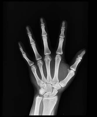
X-Rays
Introduction
X rays discovered by William Roentgen in 1895 honoured by Nobel Prize in 1901. Basically are high energy photons (1- 100Kev) or electro-magnetic radiation, having a very short wavelength of the order of 1A⁰. Typical x-ray wavelength lies between the range of
1 A⁰ and 100A⁰. Accordingly they are classified into hard X-rays and soft X-rays.
Uses of X rays are multifold. Usually they are used in surgery, radiotherapy, engineering, industry, detective department, scientific research and so many other areas. Hence, the name X-rays is popularly known even to a common man.
Uses of X rays are multifold. Usually they are used in surgery, radiotherapy, engineering, industry, detective department, scientific research and so many other areas. Hence, the name X-rays is popularly known even to a common man.
Production Of X-RAYS
The modern X-ray tube was designed by Dr.Coolidge and is known after his name as coolidge tube. It is widely used for commercial and medical purposes.
Construction and working of Coolidge Tube
The construction of coolidge tube to produce X-rays is schematically shown below.
The cathode C consists of a tungsten filament heated by passing a current through it. It then emits a large number of electrons called thermions which travel towards the anti-cathode A, also called the target with a velocity depending upon the potential difference applied between A and C.
By surrounding the cathode with a molybdenum shield M, maintained at a negative potential with respect to the filament circuit, the rays can be focused to a fine spot on A, as without the shield the electrons would fly away in all directions. The tube is completely exhausted to the no-discharge stage, so that no ionisatikn can take place.
A current of 30 mA at a voltage of 100,000 volts is used; the high voltage is obtained from a step-up transformer. The tube acts as its own rectifire, unless the anode attains such a high temperature that it begins to emit electrons. In large plants, the current from the transformers is rectified by thermoionic rectifires and high voltage capacitors.
The anti-cathode in a Coolidge tube must have the following characteristics:
- High atomic weight, to produce hard X rays.
- High melting point to withstand the high temperature developed, as most of the energy of the impinging electron is converted into heat.
- High thermal conductivity, to get rid of the heat produced.
- Low vapour pressure at high temperatures.
Tantalum, platinum and tungsten fulfil these requirements, but tungsten is the best and is widely used. In actual practice, a heavy block of this metal is mounted on a thick copper rod and the face is sloped at about 45⁰ to the electron beam. The heat generated by the impact of electrons is removed by special devices. In some cases, the anode is water-cooled.
Control Of Intensity and Quality
- The higher the potential difference between the cathode and the anti-cathode, the greater is the velocity of electrons emitted. The electrons possessing greater energy produce more penetrating Express. The quality of X-rays, therefore, where is with the difference of potential between the cathode and the anti-cathode.
- The number of electrons given out by the filament is proportional to its temperature, which can be adjusted by wearing the current in the filament circuit. The intensity of X-rays, therefore, where is with the filament current. Thus separate control of quality and intensity can be achieved.
Properties of X rays
- They are not deflected by electric and magnetic fields. This property distinguishes them from the cathode rays and indicates that the X-rays do not consist of charged particles.
- They are highly penetrating and can pass through many solids, which are Opaque to ordinary light, example, wood, flesh etc. The transparency depends upon the the density of the material. The density of substance the less transparent it is to X-rays. A sheet of lead 1 millimetre thick, absorbs these rays, where as the rays can pass through the same thickness of aluminium.
- The penetrating power of X-Rays depends upon: (a) The applied potential difference (b) The atomic weight of the material of the anti-cathode. The greater the potential difference and higher the atomic weight, the most penetrating are the X-rays produced. The X-rays having high penetrating power are known as hard X rays and those with low penetrating power are known as soft X-rays.
- They cause fluorescence in many substances, example, barium platino-cyanide, cadmium tungstate, zinc sulphide, etc.
- They produce of photochemical reaction and effect of photographic plate and they are even more effective than light.
- They ionise a gas and also eject electrons from certain metals on which they fall(For Photo electric effect click here)
- They are propagated in straight lines with the velocity of light.
- Like light, X-Rays consist of Electromagnetic waves of very short wavelength and show reflection, refraction, interference, diffraction and polarisation in a similar way. These effects can be demonstrated by suitable arrangements.
- When X-Rays are incident on matter, they gave rise to a complex phenomenon known as secondary radiations, which consist of three different types of rays.
- They have a destructive effect on living tissues. Exposure of human body to X-Rays causes reddening of the skin and surface sores.
Types of Rays after the incidence of X-rays on matter
- SCATTERED X-Rays: They are practically of the same nature and wavelength as the original or the primary rays. Their properties do not depend upon the nature of the scattering substance.
- Corpuscular Rays: They consist of fast moving electrons produced by the photoelectric process. Their properties are independent of nature of the scattering substance, but depends upon the quality of primary X-rays.
- Characteristic X-Rays: The wavelength of the characteristic X-Rays is equal to or less than the primary X-Rays, but it is characteristic of the substance upon which the primary X-Rays are incident and is independent of the wavelength of the primary X-Rays.
Uses of X-Rays
1) Surgery - X-Rays can pass through flesh and not through bones. They are of extensive use in Surgery to detect fractures, foreign bodies diseased organs etc. The patient is made to stand between an X-Ray source and a fluorescent screen.
The luminosity of the screen depends upon the stopping power of different parts of the body. Thus a depe shadow of the bones and a light shadow of the flesh will be obtained. It is called radiograph. If the various parts do not show sufficient contrast, artificial means are adopted e.g., in radiographing the portions of the alimentary tract, the patient is fed with a meal of barium sulphate.
2) Radiotherapy - X-Rays kill the diseased tissues of the body. Hence they are used to cure intractable skin diseases, malignant tumours etc. If the affected part is superficial soft rays are applied and for deep seated organs hard rays are used.
3) Engineering - Because of their high penetrating power, they are used to investigate the structure of metals, testing of castings for flaws and gas pockets, causes of weakness in structures and to detect cracks and blow holes in metal plates.
The method has the advantage that unlike some mechanical tests, it does not destroy the piece. Moreover, it often suggests alterations i mould design and heat treatment to avoid a recurrence of the detected faults.
4) Industry - X-Rays are employed in industries to detect defects in motor tyres, golf and tennis balls, wood and wireless valves. They are used in testing the uniformity of insulating material and for detecting the presence of pearls im oysters.
5) Detective Department - X-Rays are used at customs post for detection of Contraband goods like explosives, opium, etc. Concealed in leather or wooden cases, in examining of parcels without opening them and in the detection of precious metals like gold and silver in the body of smugglers. They are also used in distinguishing real diamonds, gems and pearls from artificial ones, pure ghee from vegetable ghee and real documents from forged ones.
6) Scientific Research - X-Rays have even been employed to investigate the structure of crystals, structure and properties of atoms and arrangement of atoms and molecules in matter.
Click on the sidemenu and follow the blog!
Click on subscribe button for email subscription.
Share your feedback down in the comment box.
Click on subscribe button for email subscription.
Share your feedback down in the comment box.
Thanks for reading!
Our facebook page- KnowPhysics facebook page
Our Quora space - KnowPhysics Quora
Our facebook page- KnowPhysics facebook page
Our Quora space - KnowPhysics Quora


Comments
Post a Comment
If you have any doubts or suggestions please kindly share.